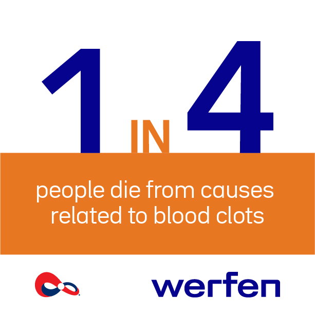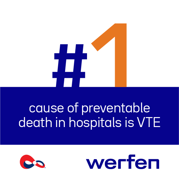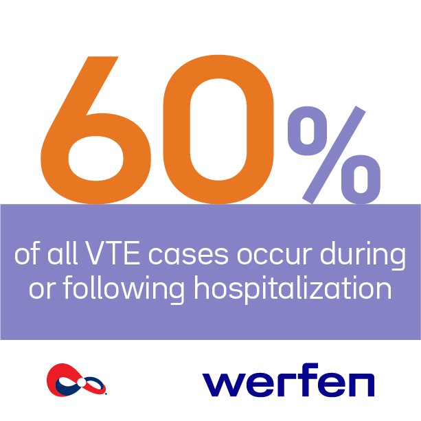
Thrombosis
Awareness
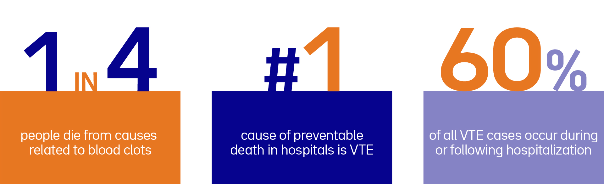
Werfen is committed to Powering Patient Care and increase global awareness of thrombosis to enhance patient outcomes.

At Werfen, our dedication to Powering Patient Care is reflected by our efforts to deepen clinical understanding of thrombosis and support better clinical outcomes.
In this episode we highlight Splanchnic Venous Thrombosis (SVT), a rare but significant subset of venous thromboembolism characterized by clot formation in the veins draining abdominal organs.
SVT represents a heterogeneous group of disorders affecting the portal, mesenteric, splenic, and hepatic veins. While commonly linked to liver cirrhosis and malignancy, SVT also can also occur in non-cirrhotic, non-neoplastic patients. Its clinical relevance stems from complications such as intestinal ischemia, portal hypertension, and potential underlying systemic disorders, including malignancies, nephrotic syndrome, thrombophilia, or inflammatory diseases.
The development of SVT follows the principles of Virchow’s triad: venous stasis, endothelial injury, and hypercoagulability. The unique anatomy and physiology of the splanchnic circulation, characterized by slower flow and exposure to inflammatory stimuli, make it particularly vulnerable to clot formation.
Compared to deep vein thrombosis and pulmonary embolism, SVT is less common and often underdiagnosed. It may be symptomatic or incidental and it typically affects the portal vein and superior mesenteric vein, with variable severity.
Although its epidemiological profile remains under-characterized, SVT is more frequently observed in advanced liver cancer. In patients without liver disease, SVT is rare and typically associated with systemic prothrombotic states, which influence both prognosis and treatment strategies.
SVT can be provoked by a variety of transient and persistent risk factors. Transient causes include abdominal surgery, inflammation, infection, pregnancy, or hormone therapy. Persistent risk factors include liver cirrhosis, thrombophilia, solid tumors, autoimmune disorders and myeloproliferative neoplasms, such as polycythemia vera or essential thrombocythemia.
The symptoms of SVT may vary depending on the location and extent of thrombosis. Acute cases can cause abdominal pain, ascites, or gastrointestinal bleeding, while chronic SVT may remain silent.
Due to the limited reliability of D-dimer testing, imaging techniques are central to diagnosis. In cases without clear etiology, laboratory evaluation for thrombophilia and genetic mutations is recommended.
Treatment of SVT requires careful consideration of clot progression versus bleeding risk, particularly in patients with cirrhosis or varices. Anticoagulant options include low molecular weight heparin, warfarin, and direct oral anticoagulants. Therapy decisions depend on the cause, clot burden, and bleeding risk. Despite treatment, recurrence remains a concern, highlighting the importance of personalized risk assessment.
SVT is a complex condition that requires a personalized approach to diagnosis and treatment. Advances in imaging and genetic profiling have enabled earlier and more precise detection, facilitating proactive management and reducing the risk of severe complications. The integration of personalized care and innovative technologies is reshaping the clinical approach to SVT, ultimately enhancing long-term patient outcomes.