Thrombosis
Awareness
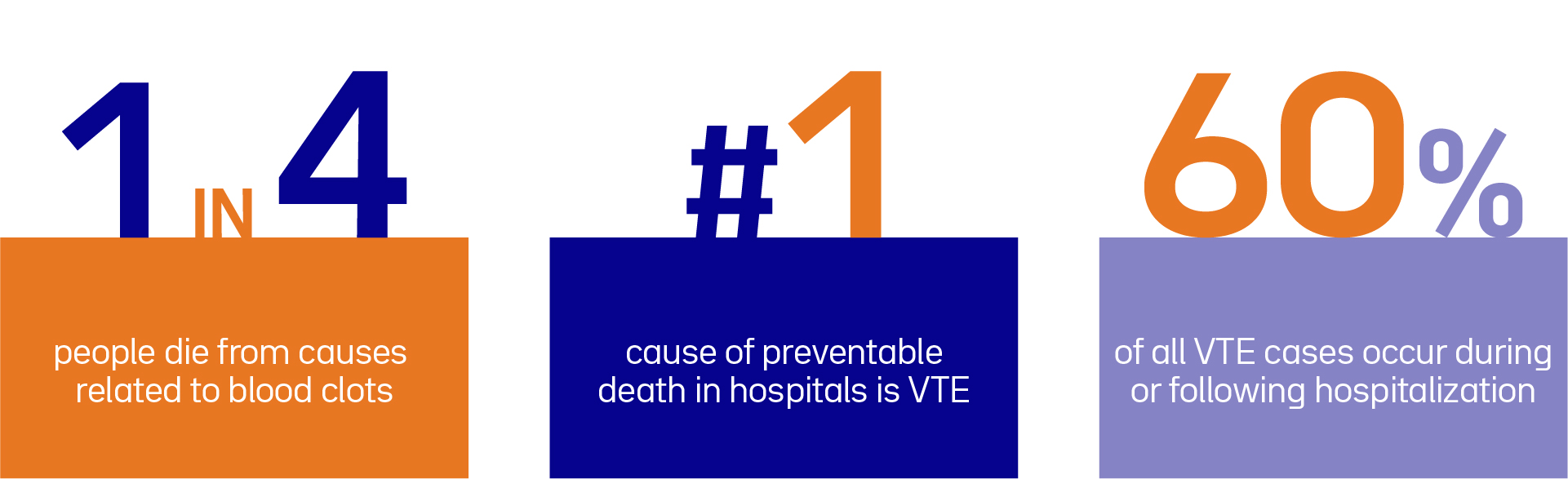
Werfen is committed to Powering Patient Care and increase global awareness of thrombosis to enhance patient outcomes.
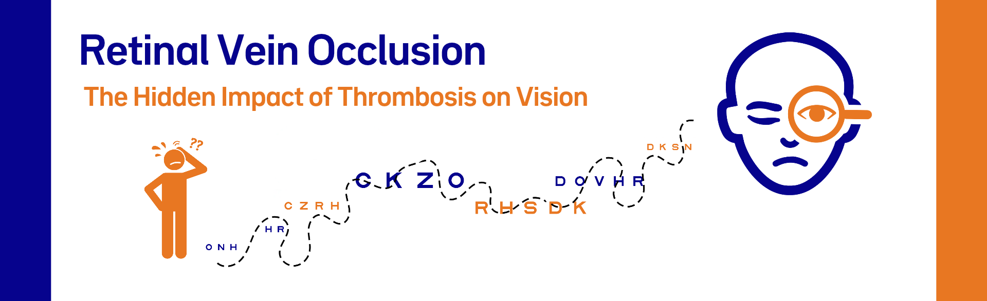
In Episode 26, we take a closer look at retinal vein occlusion, a common vascular condition that significantly contributes to visual impairment across the globe. Join us in spreading awareness on thrombosis and advancing patient care!
At Werfen, we are committed to advancing patient care through educational initiatives that increase awareness of thrombotic disorders and their clinical consequences. In this episode, we explore retinal vein occlusion (RVO), one of the most common vascular diseases of the eye and a major cause of visual impairment worldwide.
Retinal vein occlusion occurs when a blood clot or vessel compression blocks a retinal vein, trapping blood and fluid in the retina and leading to venous congestion, retinal hemorrhages, edema, and retinal ischemia. It can affect either the central retinal vein (CRVO) or one of its branches (BRVO), with BRVO being more common.
The development of RVO is influenced by several systemic conditions, including hypertension, diabetes mellitus, hyperlipidemia, cardiovascular disease, smoking, and advanced age. Although inherited thrombophilic disorders have been investigated, routine screening for these conditions is generally not recommended.
The pathogenesis of retinal vein occlusion is multifactorial and differs between central and branch forms. CRVO is primarily associated with mechanical narrowing of the vein’s lumen, while BRVO is typically due to a combination of vein compression at the arteriovenous junction, vessel wall degeneration, and abnormal hematological factors. RVO-associated ischemia and inflammation lead to upregulation of vascular endothelial growth factor (VEGF) and expression of inflammatory cytokines.
RVO typically presents as a sudden, painless reduction in vision in one eye, with severity depending on the location and extent of the blockage. Blindness is primarily driven by complications such as macular edema and retinal ischemia. While macular edema remains the leading cause of vision loss, retinal ischemia can drive pathological neovascularization, potentially resulting in vitreous hemorrhage and neovascular glaucoma.
RVO is the second leading cause of vision loss after diabetic retinopathy (DR). While both disorders affect the retinal vasculature, their mechanisms differ. RVO results from an acute venous blockage, causing sudden vision loss, usually in one eye. In contrast, DR develops progressively as chronic hyperglycemia damages retinal capillaries and is often affecting both eyes over time.
Fundus photography remains a cornerstone in the diagnosis of RVO, complemented by more advanced imaging modalities such as optical coherence tomography and fluorescein angiography. AI and deep learning models are now transforming the interpretation of retinal images and clinical data, enabling earlier detection and improved risk stratification.
Current treatment strategies primarily target complications rather than preventing disease progression. Intravitreal anti-VEGF therapy remains the standard of care for macular edema, while laser photocoagulation is still used in some cases to manage neovascular complications. Corticosteroids may benefit selected patients, whereas emerging dual inhibitor therapy aims to extend treatment durability and reduce injection frequency.
Retinal vein occlusion is a common and potentially sight-threatening vascular condition. Early diagnosis, prompt treatment, and comprehensive management of associated systemic diseases are vital to preserving vision and reducing complications. Continued advances in retinal imaging and targeted therapies are improving outcomes and offering new opportunities for personalized patient care.